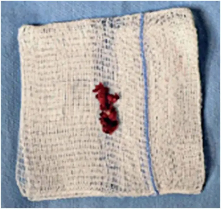
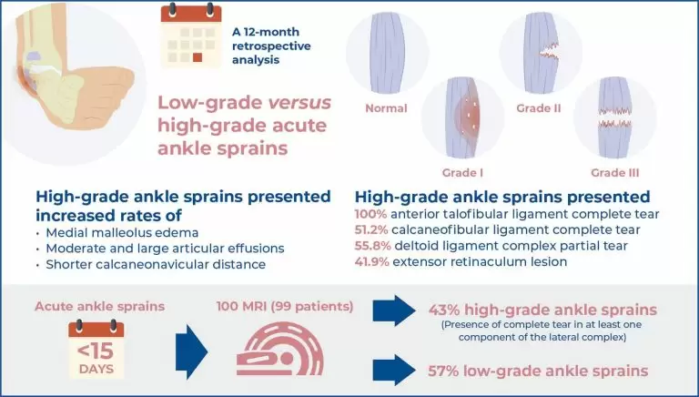
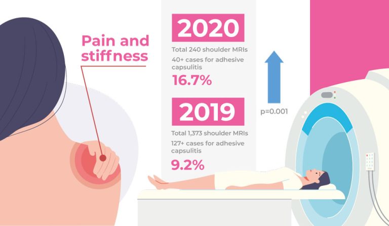
10/Jun/2025
Uretero-fallopian fistula after gynecological surgery for endometriosis: a case report
DOI: 10.31744/einstein_journal/2025RC1294
ABSTRACT The increasing use of videolaparoscopy for the complete excision of endometriotic lesions and restoration of pelvic anatomy has helped improve long-term outcomes. However, risks remain, including rare but significant complications such as salpingoureteral fistulas. Early diagnosis using imaging methods enables adequate treatment and reduces the risk of new complications. In this report, we present a case of uretero-fallopian fistula following gynecological surgery for the resection of endometriotic lesions, with intraoperative inadvertent ureteral injury. This case report aimed to highlight […]
Keywords: Fallopian tubes; Gynecologic surgical procedures; Laparoscopy; Magnetic resonance imaging; Postoperative complications; Tomography, x-ray computed; Urinary fistula; Video recording; Wounds and injuries
28/Apr/2025
Quality assessment of clinical practice in neuro-oncology
DOI: 10.31744/einstein_journal/2025AO1343
Highlights ■ The molecular test according to the WHO classification of CNS tumors (gliomas) was performed in 100% (172) of patients. ■ Education on chemotherapy and informed consent for patients with brain tumors was carried out for 100% (130) of the patients who had an indication for chemotherapy treatment. ■ Approximately 87% of the patients with gliomas underwent postoperative cranial MRI. ■ A multidisciplinary approach, including molecular testing, was used for 35% of patients with glioma. ■ Thromboembolic events after […]
Keywords: Brain neoplasms; Central nervous system neoplasms; Glioma; Length of stay; Magnetic resonance imaging; Meningioma; Patient outcome assessment; Postoperative complications; Quality improvement; Quality Indicators, Health Care
21/Nov/2024
Chronic spontaneous non-aneurysmal aortic rupture treated with endovascular surgery
DOI: 10.31744/einstein_journal/2024RC1113
ABSTRACT Spontaneous non-aneurysmal aortic rupture is rare and is usually attributed to penetrating aortic ulcers, infections, tumor infiltrations, or inflammatory and collagen diseases. Chronic rupture is infrequent but extremely rare in non-aneurysmal aortas, which makes diagnosis difficult because the absence of an aneurysm can mislead the physician to rule out rupture. Here, we describe the case of an 85-year-old male, who was undergoing oncological investigation for weight loss, inappetence, and back pain. Computed tomography and magnetic resonance imaging performed 3 […]
Keywords: Aged, 80 and over; Aneurysm; Aneurysm, false; Angiography; Aorta, abdominal; Endovascular procedures; Intensive care units; Magnetic resonance imaging; Patient discharge; Penetrating atherosclerotic ulcer; Prostheses and implants; Rupture; spontaneous; Tomography, x-ray computed
06/Jun/2024
Effect of transcranial direct current stimulation in the initial weeks post-stroke: a pilot randomized study
DOI: 10.31744/einstein_journal/2024AO0450
Highlights Differences ≥9 points in Fugl–Meyer Assessment scores: sham>active. Connectivity measures: active>sham. ABSTRACT Objective: This study aimed at assessing the alterations in upper limb motor impairment and connectivity between motor areas following the post-stroke delivery of cathodal transcranial direct current stimulation sessions. Methods: Modifications in the Fugl-Meyer Assessment scores, connectivity between the primary motor cortex of the unaffected and affected hemispheres, and between the primary motor and premotor cortices of the unaffected hemisphere were compared prior to and following six […]
Keywords: Connectome; Magnetic resonance imaging; Motor cortex; Neurological rehabilitation; Stroke; Transcranial direct current stimulation
15/Sep/2023
Acute ankle injuries: association between sprain severity and ancillary findings
DOI: 10.31744/einstein_journal/2023AO0162
Highlights Participants presenting acute ankle sprains (<15 days) were divided into low-grade versus high-grade sprain, according to the presence of a complete tear in at least one component of lateral ligament complex. High-grade ankle sprains group presented increased rates of medial malleolus bone bruise, deltoid ligament tears, extensor retinaculum lesions, and articular effusion. Thecalcaneonaviculardistancewasstatisticallyshorterin patients with high-grade sprains (median, 3.0mm) when compared to those with low-grade sprains (median, 4.0mm). INTRODUCTION Ankle fractures and sprains are extremely common injuries, among the […]
Keywords: Ankle/injuries; Fractures, avulsion; Ligaments/ injuries; Magnetic resonance imaging; Sprains and strains
21/Nov/2022
Incidence of adhesive capsulitis of the shoulder during the beginning of the COVID-19 pandemic
DOI: 10.31744/einstein_journal/2022AE0163
ABSTRACT Objective Adhesive capsulitis is an inflammatory disease of the joint capsule, clinically manifested as pain, stiffness, and dysfunction of the shoulder. We subjectively observed an increased incidence of adhesive capsulitis, and raised the hypothesis that adhesive capsulitis was more frequent in magnetic resonance imaging examinations performed during the COVID-19 pandemic as compared with examinations prior to this period. Methods Data from medical records and magnetic resonance imaging of the shoulder presenting typical imaging findings of adhesive capsulitis, performed in […]
Keywords: Betacoronavirus; Bursitis; Coronavirus infections; COVID-19; Magnetic resonance imaging; SARS-CoV-2; Shoulder
21/Nov/2022
Prostate imaging features on magnetic resonance imaging of young patients
DOI: 10.31744/einstein_journal/2022AO0024
ABSTRACT Objective To identify magnetic resonance imaging findings of the prostate in young adults, including symptomatic and asymptomatic patients. The aim of this study is to evaluate the main aspects of prostate imaging in young patients. Methods A total of 102 patients under 40 years of age, who underwent prostate magnetic resonance imaging between January 2016 and January 2019, were included in this study. The patients were divided into two groups: symptomatic for prostatitis (Group 1) and asymptomatic (Group 2). […]
Keywords: Magnetic resonance imaging; Prostate; Prostatic neoplasms
08/Jul/2022
FAST breast magnetic resonance imaging: a new approach for breast cancer screening?
DOI: 10.31744/einstein_journal/2022AO0073
ABSTRACT Objective To develop an abbreviated breast magnetic resonance imaging protocol (FAST) and to compare it with the complete protocol (FULL) to determine its diagnostic accuracy for detecting malignant or suspicious lesions (BI-RADS 4, 5 and 6) and the time required for image interpretation using BI-RADS categorization. Methods Retrospective study with 100 consecutive women who underwent breast magnetic resonance imaging between January and February 2014. All patients were submitted to a complete breast magnetic resonance imaging protocol, which was then […]
Keywords: Breast; FAST; Magnetic resonance imaging; Mass screening
18/May/2022
T2-weighted imaging hypointensity in an ovarian lesion: is it a benign finding?
DOI: 10.31744/einstein_journal/2022AO6851
ABSTRACT Objective To evaluate whether the presence of a hypointense signal at T2-weighted imaging in a solid ovarian lesion on magnetic resonance imaging is a predictor of stability and benignity. Methods This is a single center study, prospectively read with retrospective acquired data. The database was searched for patients who underwent magnetic resonance imaging between January 2008 and October 2019 and whose reports mentioned solid ovarian lesions with low signal on T2-weighted imaging. A total of 47 nodules were included. […]
Keywords: Algorithms; Magnetic resonance imaging; Magnetic resonance spectroscopy; Ovarian neoplasms
07/Apr/2022
Degenerative injuries of the metatarsophalangeal plantar plate on magnetic resonance imaging: a new perspective
DOI: 10.31744/einstein_journal/2022AO6543
ABSTRACT Objective The magnetic resonance imaging diagnostic criteria for a complete tear of metatarsophalangeal plantar plate are well-established. However, more subtle abnormalities can also occur and be a source of pain. The objective of this study is to determine the prevalence of degenerative plantar plate injuries in patients with metatarsalgia who underwent forefoot magnetic resonance imaging and describe the main abnormalities found. The hypothesis is that mild capsular fibrosis will have high sensitivity but low specificity for plantar plate degenerative […]
Keywords: Joint instability; Magnetic resonance imaging; Metatarsalgia; Metatarsophalangeal joint; Plantar plate; Rupture