26/Nov/2024
APENDIC-RADS: an ultrasound reporting system for the diagnosis of acute appendicitis
DOI: 10.31744/einstein_journal/2024AO1164
Highlights ■ Imaging plays a fundamental role in the diagnosis of acute appendicitis. ■ The study highlights ultrasound’s effectiveness in diagnosing appendicitis. ■ APPENDIC-RADS efficiently standardizes the ultrasound evaluation of appendicitis. ■ Using APPENDIC-RADS may enhance communication and optimize medical practices. ABSTRACT Objective: Abdominal ultrasonography is widely used to evaluate suspected cases of appendicitis. Objective descriptions of the direct and indirect signs of appendicitis result in varied assessments of its likelihood. This study introduces the Appendix Imaging Reporting and Data […]
Keywords: Appendicitis; Emergency medicine; Interdisciplinary communication; Ultrasonography
21/Nov/2024
Point-of-care ultrasonography for diagnosis of purulent pericarditis postappendectomy: a case report
DOI: 10.31744/einstein_journal/2024RC0962
ABSTRACT Purulent pericarditis is rare condition in the modern era of antibiotics. However, it is a serious condition as it has an accelerated progression and is difficult to diagnose due to its nonspecific clinical presentation, resulting in high mortality. Herein, we present a case in which a 36-year-old male patient with otherwise unremarkable medical history developed abdominal sepsis complicated by purulent pericarditis post-appendectomy. While the initial clinical presentation was not compatible with the classic signs of purulent pericarditis, the diagnosis […]
Keywords: Appendectomy; Critical care; Pericardial effusion; Pericarditis; Suppuration; Ultrasonography
20/May/2024
Lessons from the pandemic and the value of a structured system of ultrasonographic findings in the diagnosis of COVID-19 pulmonary manifestations
einstein (São Paulo). 20/May/2024;22:eAE0780.
View Article20/May/2024
Lessons from the pandemic and the value of a structured system of ultrasonographic findings in the diagnosis of COVID-19 pulmonary manifestations
DOI: 10.31744/einstein_journal/2024AE0780
Highlights Lung ultrasound enables precise diagnosis and ongoing monitoring of the disease. Ultrasound is an effective tool for assessing pulmonary findings in COVID-19. Structured reports enhance communication and are easily reproducible. ABSTRACT Objective: We aimed to share our experience in implementing a structured system for COVID-19 lung findings, elucidating key aspects of the lung ultrasound score to facilitate its standardized clinical use beyond the pandemic scenario. Methods: Using a scoring system to classify the extent of lung involvement, we retrospectively […]
Keywords: Clinical decision-making; Coronavirus infections; COVID-19; Lung disease; Organ dysfunction; SARS-CoV-2; Ultrasonography
09/Nov/2022
Sonographic signs of deep infiltrative endometriosis among women submitted to routine transvaginal sonography: clinical and imaging aspects
DOI: 10.31744/einstein_journal/2022AO0086
ABSTRACT Objective To evaluate the prevalence of sonographic signs suggestive of deep infiltrative endometriosis and endometriomas in patients referred for transvaginal sonography as part of a routine annual gynecological evaluation. We also describe the clinical and imaging aspects associated with the incidental findings of endometriosis. Methods This was a retrospective observational study including women (n=339; aged 18-56 years) referred for transvaginal sonography as part of a routine gynecological evaluation (without clinical suspicion of endometriosis). Patients were asked about their symptoms. […]
Keywords: Endometrial neoplasms; Endometriosis; Prevalence; Ultrasonography
28/Jun/2022
Ultrasonographic evaluation of cecal appendix diameter in pediatric population
DOI: 10.31744/einstein_journal/2022AO6935
ABSTRACT Objective To stratify ultrasound samples in a pediatric population undergoing evaluation for acute appendicitis to examine the variability in cecal appendix diameter, in different age groups, and to determine whether there is a prevalent value for each age group. Methods A retrospective cross-sectional study with 196 children aged 0 to 15 years. Data were extracted from reports of ultrasound examinations carried out between 2008 and 2015. Children with sonographic diagnosis of appendicitis or other signs of periappendiceal inflammation were […]
Keywords: Appendicitis/diagnostic imaging; Appendix; Child; Ultrasonography
06/May/2022
Ultrasound classification of thyroid nodules: does size matter?
DOI: 10.31744/einstein_journal/2022AO6747
ABSTRACT Objective To determine whether the size of thyroid nodules in ACR-TIRADS ultrasound categories 3 and 4 is correlated with the Bethesda cytopathology classification. Methods Thyroid nodules (566) subclassified as ACR-TIRADS 3 or 4 were divided into three size categories according to American Thyroid Association guidelines. The frequency of different Bethesda categories in each size range within ACR-TIRADS 3 and 4 classifications was analyzed. Results Most nodules in both ACR-TIRADS classifications fell in the Bethesda 2 category, regardless of size […]
Keywords: ACR-TIRADS; Bethesda; Malignance; Thyroid gland nodule; Thyroid gland/pathology; Ultrasonography
07/Apr/2022
Testicular volume and Tanner stage: determinant factors for testicular torsion
DOI: 10.31744/einstein_journal/2022AO6605
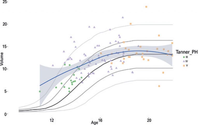
ABSTRACT Objective To assess testicular volumes and sexual maturation in patients with testicular torsion. Methods A retrospective analysis of consecutively treated patients with testicular torsion between 2016 and 2018. Age, pubic hair staging (Tanner), and by ultrasonography, volume of the unaffected testis (in cubic centimeters) were evaluated either immediately before surgery or at the first postoperative visit. Patients with previous testicular disease, such as cryptorchidism, or with no records of testicular volume were excluded. The analysis included descriptive statistics and […]
Keywords: Ischemia; Sexual maturation; Spermatic cord torsion; Testis/diagnostic imaging; Testis/growth & development; Ultrasonography
26/Nov/2020
Caecal appendix lipomatosis in a pregnant patient mimicking acute appendicitis
DOI: 10.31744/einstein_journal/2020RC5415
ABSTRACT A 34-years-old pregnant woman admitted in the emergency unit complaining about worsening right iliac fossa pain for 2 days. Acute appendicitis was the suspected diagnosis. Laboratory exams were ordered and results were within normal limits for infectious and inflammatory aspects. Ultrasound scan revealed a pregnancy in course without alterations and a thickness of the appendix wall without inflammatory signs in the surrounding tissue. Because the suspicion of acute appendicitis remained, a magnetic resonance was done and confirmed the diagnosis […]
Keywords: Appendicitis; Diagnosis, differential; Lipomatosis; Magnetic resonance imaging; Pregnant Women; Ultrasonography
06/Nov/2020
A comparative study of ultrasound and cross-sectional imaging for detection of small renal masses: anatomic factors and radiologist’s experience
DOI: 10.31744/einstein_journal/2020AO5576
ABSTRACT Objective: To evaluate anatomic factors and radiologist’s experience in the detection of solid renal masses on ultrasonography. Methods: We searched for solid renal masses diagnosed on cross-sectional imaging from 2007 to 2017 that also had previous ultrasonography from the past 6 months. The following features were evaluated: nodule size, laterality, location and growth pattern, patient body mass index and radiologist’s experience in ultrasound. In surgically resected cases, pathologic reports were evaluated. Unpaired t test and χ2 test were used […]
Keywords: Diagnostic imaging; kidney neoplasms/diagnostic, imaging; Multidetector computed tomography; Ultrasonography
28/Jan/2020
Prostate volume measurement by multiparametric magnetic resonance and transrectal ultrasound: comparison with surgical specimen weight
DOI: 10.31744/einstein_journal/2020AO4662
ABSTRACT Objective To assess accuracy of multiparametric magnetic resonance of the prostate to estimate gland volume, comparing the results with transrectal ultrasound and surgical specimen. Methods A retrospective study of 85 patients who underwent multiparametric magnetic resonance and transrectal ultrasound (for fusion image-guided biopsy) before radical prostatectomy. Prostate measurements were obtained from magnetic resonance axial and sagittal T2-weighted images and ultrasound; the prostate volume was determined using the ellipsoid formula. The results were compared with the surgical specimen weight. Maximum […]
Keywords: Magnetic resonance imaging; Magnetic resonance spectroscopy; Prostatectomy; Prostatic diseases; Ultrasonography