20/Sep/2024
Cluster classification of a Brazilian gastric cancer cohort reveals remarkable populational differences in normal p53 rate
einstein (São Paulo). 20/Sep/2024;22:eAO0508.
View Article20/Sep/2024
Cluster classification of a Brazilian gastric cancer cohort reveals remarkable populational differences in normal p53 rate
DOI: 10.31744/einstein_journal/2024AO0508
ABSTRACT In Brief Queiroz et al. showed that the application of cluster methodology for classifying gastric cancer is suitable and efficient within a Brazilian cohort, which is known for its population heterogeneity. The study highlighted the potential utilization of this method within public health services due to its low-cost, presenting a viable means to improve the diagnosis and prognosis of gastric cancer. Highlights Our Brazilian cohort with gastric cancer has a distinct distribution between mutated and normal p53. New genetic […]
Keywords: Adenocarcinoma; Biomarkers; Cluster analysis; Gastric neoplasms; Genes p53; Genetic heterogeneity; Immunohistochemistry
14/Feb/2019
Clinicopathological and immunohistochemical analysis of spindle cell squamous cell carcinoma of the tongue: a rare case
einstein (São Paulo). 14/Feb/2019;17(1):eRC4610.
View Article14/Feb/2019
Clinicopathological and immunohistochemical analysis of spindle cell squamous cell carcinoma of the tongue: a rare case
DOI: 10.31744/einstein_journal/2019RC4610
ABSTRACT Spindle cell squamous cell carcinoma of the tongue is a rare variant of squamous cell carcinoma. This paper reports the case of a spindle cell squamous cell carcinoma of the tongue, in a 64-year-old male patient, and presents a review of the etiopathogenesis, clinicopathological and immunohistochemical features and treatment of the malignancy. The patient presented for evaluation of a painful swelling on his tongue. Extraoral examination revealed palpable submandibular and superior cervical lymph nodes. Based on the presumptive diagnoses […]
Keywords: Carcinoma, squamous cell; Immunohistochemistry; Mouth/pathology; Tongue neoplasms/diagnosis
03/May/2018
Clinical and immunohistochemical study of melanotic neuroectodermal tumor of infancy in the maxilla
einstein (São Paulo). 03/May/2018;16(2):eRC4025.
View Article03/May/2018
Clinical and immunohistochemical study of melanotic neuroectodermal tumor of infancy in the maxilla
DOI: 10.1590/S1679-45082018RC4025
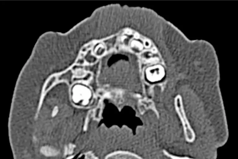
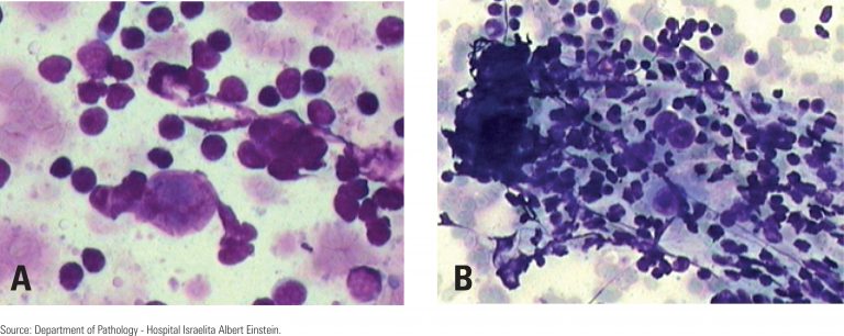
ABSTRACT Melanotic neuroectodermal tumor of infancy is a rare and fast-growing neoplasm. In this study, we describe the case of a 6-month-old female patient, who presented swelling in the anterior maxilla. Tomographic reconstruction showed an unilocular hypodense and expansive area associated with the upper right central primary incisor. The presumptive diagnoses were dentigerous cyst, adenomatoid odontogenic tumor, melanotic neuroectodermal tumor of infancy and rhabdomyosarcoma, and an incisional biopsy was performed. Microscopically, the lesion revealed a biphasic cell population, consisting of […]
Keywords: Case reports; Child; Immunohistochemistry; Maxillary neoplasms/diagnosis; Microscopy; Neuroectodermal tumor, melanotic/diagnosis
01/Apr/2016
The destruction complex of beta-catenin in colorectal carcinoma and colonic adenoma
DOI: 10.1590/S1679-45082016AO3678
ABSTRACT Objective To evaluate the destruction complex of beta-catenin by the expression of the proteins beta-catetenin, adenomatous polyposis coli, GSK3β, axin and ubiquitin in colorectal carcinoma and colonic adenoma. Methods Tissue samples from 64 patients with colorectal carcinoma and 53 patients with colonic adenoma were analyzed. Tissue microarray blocks and slides were prepared and subjected to immunohistochemistry with polyclonal antibodies in carcinoma, adjacent non-neoplastic mucosa, and adenoma tissues. The immunoreactivity was evaluated by the percentage of positive stained cells and […]
Keywords: Adenoma; Axin protein; beta Catenin; Colorectal neoplasms; Genes, APC; Glycogen synthase; Immunohistochemistry; Ubiquitin; Wnt signaling pathway
31/Jan/2014
Immunoexpression of the COX-2, p53, and caspase-3 proteins in colorectal adenoma and non-neoplastic mucosa
einstein (São Paulo). 31/Jan/2014;11(4):456-61.
View Article31/Jan/2014
Immunoexpression of the COX-2, p53, and caspase-3 proteins in colorectal adenoma and non-neoplastic mucosa
DOI: 10.1590/S1679-45082013000400009
OBJECTIVE: To analyze the immunoexpression of the COX-2, p53, and caspase-3 proteins in colorectal adenomas and non-neoplastic mucosa. METHODS: 72 individuals were subjected to colonoscopy, which provided 50 samples of adenomas and 45 samples of non-neoplastic colorectal mucosa. The tissue samples were obtained via the tissue microarray technique and subjected to immunohistochemical analysis using primary anti-p53, anti-COX-2, and anti-caspase-3 antibodies. The positivity and intensity of the immunoreaction were classified. The analyzed variables were as follows: site of the adenomas in […]
Keywords: Adenoma; Caspase 3; Cyclooxygenase 2; Gene, p53; Immunohistochemistry; Intestine, large
01/Apr/2011
Correlation between flow cytometry and histologic findings: ten year experience in the investigation of lymphoproliferative diseases
einstein (São Paulo). 01/Apr/2011;9(2 Pt 1):151-9.
View Article01/Apr/2011
Correlation between flow cytometry and histologic findings: ten year experience in the investigation of lymphoproliferative diseases
DOI: 10.1590/S1679-45082011AO2027
ABSTRACT Objective: To demonstrate the advantages of correlating flow cytometry immunophenotyping with the pathology/ immunohistochemistry of lymph nodes or nodules in the diagnosis of lymphoproliferative diseases. Methods: A retrospective study was carried out of 157 biopsy or fine-needle aspiration lymph nodes/ nodule specimens taken from 142 patients, from 1999 and 2009. The specimens were simultaneously studied with fow cytometry and pathology at Hospital Israelita Albert Einstein. The specimens were prepared in hematoxylin/eosin, Giemsa, or monoclonal antibody stained slides for detecting […]
Keywords: Flow cytometry; Immunohistochemistry; Immunophenotyping; Lymphoma; Lymphoproliferative disorders/diagnosis