The journal einstein (São Paulo) – e-ISSN 2317-6385 is dedicated to dissemination of high-quality scientific content that advances our understanding of human disease with the goal of improving prevention, care, diagnostics and treatment of patients globally.
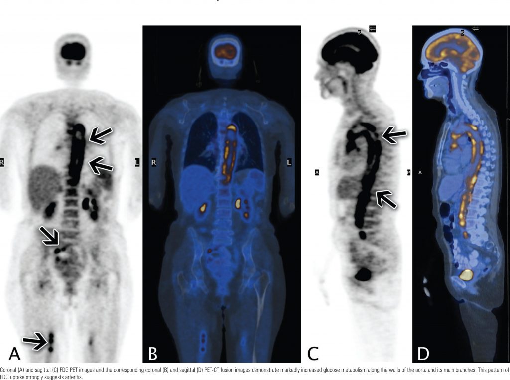
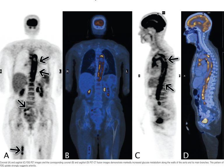
A 69-year-old Caucasian female was referred to the Imaging Department for investigation of fever of unknown origin (FUO), fatigue for seven months, and recent onset of atypical chest pain. Laboratory tests revealed elevation of serum C-reactive protein levels and erythrocyte sedimentation rate.
Positron emission tomography with computed tomography fusion (PET-CT) with F18-fluorodeoxyglucose (FDG) was performed for the evaluation of FUO by excluding malignancy. The FDG images revealed markedly diffuse and circumferential increased uptake suggesting diffuse inflammatory process along the walls of the aorta and subclavian arteries.
FlaminiRC, YamagaLYI, OsawaA, NomuraC, GuerraJCC, PasternakJ, CunhaML, et al. PET-CT findings in arteritis. Einstein (São Paulo) 2010;8(4 Pt 1):498-9. https://doi.org/10.1590/S1679-45082010AI1565
Flamini,Rodrigo de Carvalho; Yamaga,Lilian Yuri Itaya; Osawa,Akemi; Nomura,Cesar; Guerra,João Carlos de Campos; Pasternak,Jacyr; Cunha,Marcelo Livorsi da; Campos Neto,Guilherme de Carvalho; Wagner,Jairo; Funari,Marcelo Buarque de Gusmão. PET-CT findings in arteritis. Einstein (São Paulo), v. 8, n. 4 Pt 1, p. 498-499, Oct. 2010. https://doi.org/10.1590/S1679-45082010AI1565
Flamini,Rodrigo de Carvalho and Yamaga,Lilian Yuri Itaya and Osawa,Akemi and Nomura,Cesar and Guerra,João Carlos de Campos and Pasternak,Jacyr and Cunha,Marcelo Livorsi da and Campos Neto,Guilherme de Carvalho and Wagner,Jairo and Funari,Marcelo Buarque de Gusmão. PET-CT findings in arteritis. Einstein (São Paulo) [online]. 2010, vol. 8, n. 4 Pt 1, [cited 2025-09-06], pp.498-499. Available from: <https://journal.einstein.br/article/pet-ct-findings-in-arteritis/>. ISSN 1679-4508. https://doi.org/10.1590/S1679-45082010AI1565
Coronal (A) and sagittal (C) FDG PET images and the corresponding coronal (B) and sagittal (D) PET-CT fusion images demonstrate markedly increased glucose metabolism along the walls of the aorta and its main branches. This pattern of FDG uptake strongly suggests arteritis.