einstein (São Paulo). 31/Jan/2014;11(4):528-32.
Iron overload in a teenager with xerocytosis: the importance of nuclear magnetic resonance imaging
DOI: 10.1590/S1679-45082013000400022
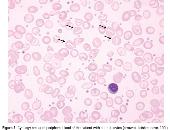
To report a case of iron overload secondary to xerocytosis, a rare disease in a teenager, diagnosed, by T2* magnetic resonance imaging. We report the case of a symptomatic patient with xerocytosis, a ferritin level of 350ng/mL and a significant cardiac iron overload. She was diagnosed by T2* magnetic resonance imaging and received chelation therapy Ektacytometric analysis confirmed the diagnosis of hereditary xerocytosis. Subsequent T2* magnetic resonance imaging demonstrated complete resolution of the iron overload in various organs, as a new echocardiography revealed a complete resolution of previous cardiac alterations. The patient remains in chelation therapy. Xerocytosis is a rare autosomal dominant genetic disorder characterized by dehydrated stomatocytosis. The patient may present with intense fatigue and iron overload. We suggest the regular use of T2* magnetic resonance imaging for the diagnosis and control of the response to iron chelation in xerocytosis, and we believe it can be used also in other hemolytic anemia requiring transfusions.