30/Jan/2024
Littoral cell angioma of the spleen: case report and literature review
DOI: 10.31744/einstein_journal/2024RC0267
ABSTRACT Littoral cell angioma is an extremely rare splenic vascular tumor originating from the cells lining the splenic red pulp sinuses. Approximately 150 cases of littoral cell angioma have been reported since 1991. Its clinical manifestation is usually asymptomatic and is mostly diagnosed as an incidental finding through abdominal imaging. Herein, we present a case of littoral cell angioma in a 41-year-old woman with no previous comorbidities, which initially presented as a nonspecific splenic lesion diagnosed on imaging in the […]
Keywords: Hemangioma; Incidental findings; Laparoscopy; Splenectomy; Splenic neoplasms; Tomography; X-ray computed
20/May/2022
Prognostic factors of worse outcome for hospitalized COVID-19 patients, with emphasis on chest computed tomography data: a retrospective study
einstein (São Paulo). 20/May/2022;20:eAO6953.
View Article20/May/2022
Prognostic factors of worse outcome for hospitalized COVID-19 patients, with emphasis on chest computed tomography data: a retrospective study
DOI: 10.31744/einstein_journal/2022AO6953
ABSTRACT Objective: To evaluate anthropometric and clinical data, muscle mass, subcutaneous fat, spine bone mineral density, extent of acute pulmonary disease related to COVID-19, quantification of pulmonary emphysema, coronary calcium, and hepatic steatosis using chest computed tomography of hospitalized patients with confirmed diagnosis of COVID-19 pneumonia and verify its association with disease severity. Methods: A total of 123 adults hospitalized due to COVID-19 pneumonia were enrolled in the present study, which evaluated the anthropometric, clinical and chest computed tomography data […]
Keywords: Coronavirus infections; COVID-19; Multidetector computed tomography; Obesity; Pneumonia; Prognosis; Tomography; X-ray computed
01/Jul/2016
Preclinical molecular imaging: development of instrumentation for translational research with small laboratory animals
einstein (São Paulo). 01/Jul/2016;14(3):408-14.
View Article01/Jul/2016
Preclinical molecular imaging: development of instrumentation for translational research with small laboratory animals
DOI: 10.1590/S1679-45082016AO3696
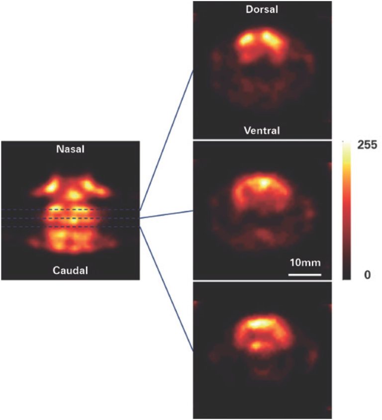
ABSTRACT Objective: To present the result of upgrading a clinical gamma-camera to be used to obtain in vivo tomographic images of small animal organs, and its application to register cardiac, renal and neurological images. Methods: An updated version of the miniSPECT upgrading device was built, which is composed of mechanical, electronic and software subsystems. The device was attached to a Discovery VH (General Electric Healthcare) gamma-camera, which was retired from the clinical service and installed at the Centro de Imagem […]
Keywords: animal; emission-computed; Likelihood functions; Models; single-photon; Tomography
21/Aug/2014
Rhino facial zygomycosis: case report
DOI: 10.1590/S1679-45082014RC2579
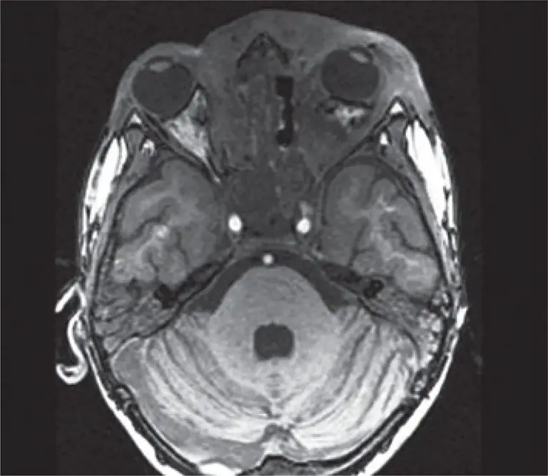
Zygomycosis is an invasive disease that affects both immunocompetent and immunocompromised, depending on the type of strain. This disease diagnosis is clinical and histopathological, and its treatment is based on antifungal therapy and surgical cleaning. This paper reports a case of a boy with invasive zygomycosis rinofacial who final treatment was successful after underwent antifungal and surgical therapies.
Keywords: Case reports; Magnetic resonance imaging; Tomography; X-ray computed; Zygomycosi/diagnosis; Zygomycosis/therapy


