The journal einstein (São Paulo) – e-ISSN 2317-6385 is dedicated to dissemination of high-quality scientific content that advances our understanding of human disease with the goal of improving prevention, care, diagnostics and treatment of patients globally.
A 63-year-old man came to the emergency room complaining of unverified fever and myalgia. Oropharyngeal material was collected for reverse transcription testing followed by polymerase chain reaction (RT-PCR) for coronavirus disease 2019 (COVID-19), and a chest radiography was performed (normal), and the patient was discharged to home isolation, due to his mild symptoms, until the test result.
[…]
Quantitative analysis in COVID-19: report of an initial experience
DalpráFAR, FonsecaEKUN, SzarfG, ChateRC. Quantitative analysis in COVID-19: report of an initial experience. Einstein (Sao Paulo). 2020;18:eAI5842. https://doi.org/10.31744/einstein_journal/2020AI5842
Dalprá,Fábio Augusto Ribeiro; Fonseca,Eduardo Kaiser Ururahy Nunes; Szarf,Gilberto; Chate,Rodrigo Caruso. Quantitative analysis in COVID-19: report of an initial experience. Einstein (Sao Paulo)., v. 18, eAI5842, Oct. 2020. https://doi.org/10.31744/einstein_journal/2020AI5842
Dalprá,F.A.R., Fonseca,E.K.U.N., Szarf,G., & Chate,R.C. (2020). Quantitative analysis in COVID-19: report of an initial experience. Einstein (Sao Paulo).,18, eAI5842. https://doi.org/10.31744/einstein_journal/2020AI5842
Dalprá,Fábio Augusto Ribeiro and Fonseca,Eduardo Kaiser Ururahy Nunes and Szarf,Gilberto and Chate,Rodrigo Caruso. Quantitative analysis in COVID-19: report of an initial experience. Einstein (Sao Paulo). [online]. 2020, vol. 18, [cited 2026-02-03], eAI5842. Available from: <https://journal.einstein.br/article/quantitative-analysis-in-covid-19-report-of-an-initial-experience/>. ISSN 1679-4508. https://doi.org/10.31744/einstein_journal/2020AI5842
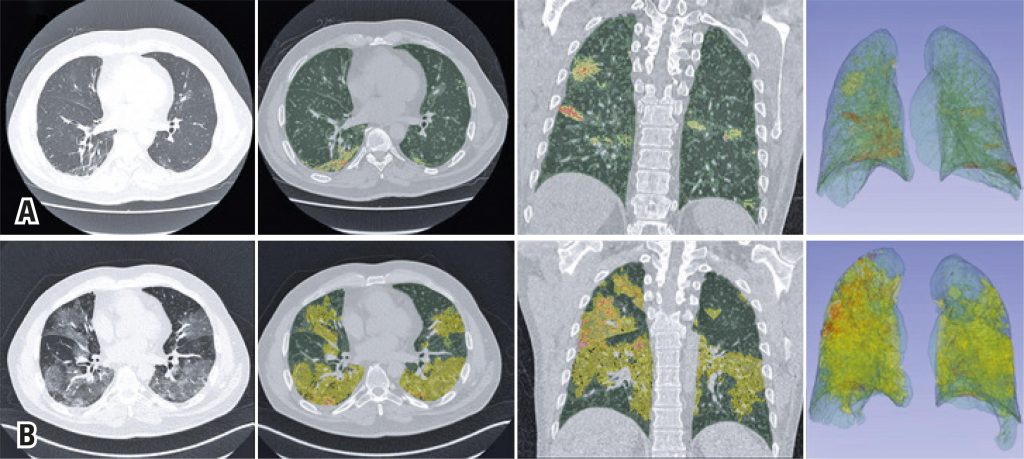
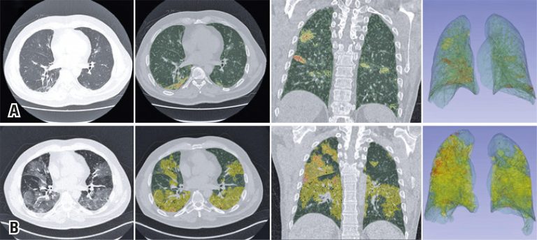
Figure 1
Chest computed tomography and superimposed 3DSlicer software quantification images. The upper series (A) show the findings when the patient returned to the emergency room, and the lower series (B) show the findings at the time of his clinical worsening. Axial sections of the chest tomography showing multifocal pulmonary ground-glass opacities predominantly peripheral and basal, more extensive in the last study, and quantitative images generated by the 3DSlicer software superimposed over the tomographic images. The areas marked in yellow show ground-glass opacities, those marked in green are areas of normal parenchyma, and those marked in orange are areas of consolidation. The extensive progression of the findings illustrates the numerical data provided
Chest computed tomography and superimposed 3DSlicer software quantification images. The upper series (A) show the findings when the patient returned to the emergency room, and the lower series (B) show the findings at the time of his clinical worsening. Axial sections of the chest tomography showing multifocal pulmonary ground-glass opacities predominantly peripheral and basal, more extensive in the last study, and quantitative images generated by the 3DSlicer software superimposed over the tomographic images. The areas marked in yellow show ground-glass opacities, those marked in green are areas of normal parenchyma, and those marked in orange are areas of consolidation. The extensive progression of the findings illustrates the numerical data provided