einstein (São Paulo). 30/Oct/2015;14(1):104-5.
A lesson learnt: retrospection in a case of pilomatricoma mimicking as parotid neoplasm
DOI: 10.1590/S1679-45082015AI3096
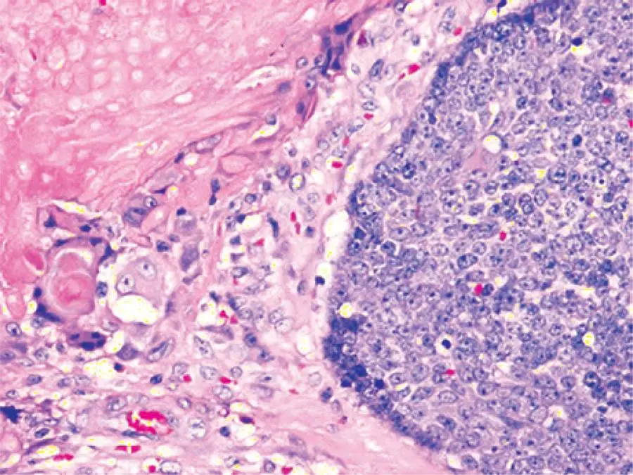
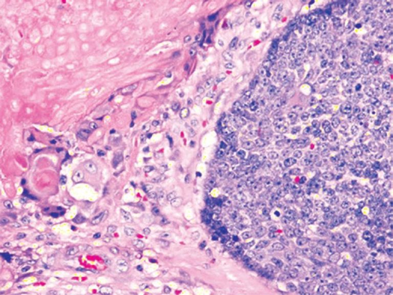
A 10-year-old girl presented with a painless, slow-growing swelling on left pre-auricular region that was noticed for 2 years (). The swelling was bosselated, non-tender, firm-to-hard on palpation, had poorly-defined margin, and measured approximately 3.5cm x 3.0cm. In addition, it had restricted mobility, and seemed to be of parotid origin, with overlying skin apparently stretched and fixed. The ultrasonography was unable to delineate the depth and confirm the involvement of the parotid, although fine needle aspiration cytology (FNAC) suggested pleomorphic adenoma. For this reason, we decided to perform superficial parotidectomy. The histopathology of the lesion diagnosed pilomatricoma, which is an uncommon, benign ectodermal tumor of dermis/subdermis. Pilomatricoma constitutes a pluripotent cell expression in the germinal matrix center of hair follicles with differentiation into cortical cells, and the main relevance of its clinical presence is its potential to be misinterpreted, resulting in unnecessary aggressive interventions. It is often a diagnosis of retrospection. Some large series reported correct pre-operative diagnosis at 1.1 to 29%.(,) A careful re-examination of pre-operative image revealed a subtle bluish tinge, which was not observed presumably because of the dark-skinned complexion, and the characteristic “tent sign”() perceptible through the already-stretched and bosselated skin surface (). Fine needle aspiration cytology is imperative for the assessment but is often erroneous() due to inadequate, non-representative sampling from a lesion with complex cell contents. In a typical example, the basophilic basaloid cells with scanty cytoplasm and indistinct borders encase the anucleated cytoplasm-rich eosinophilic “ghost/shadow cells” () with areas of keratinization, giant-cells and dystrophic calcification in the matrix. As the tumor gets older, the central “ghost cells” increase at the expense of the peripheral basophilic cells. Thus, FNAC might have more basophilic cells in the lesion in early stages while keratinocytes might predominate in later stages, so that the lesion can be misrepresented as malignant in both stages.() The problem is complicated by the fact that the parotid region is one of the most common topographic areas with involvement of head and neck pilomatricomas,(,-)which also increase the dilemma of clinicians and pathologists because pilomatricoma is seldom considered as a differential diagnosis for a mass lesion involving this region. In fact, some reports on FNAC from such lesions had suggested parotid neoplasm,() or even metastatic malignant deposits.() Because surgeons relying greatly on cytology in clinically suspected parotid tumors, such approach have resulted in more aggressive surgical treatment.(,,)
[…]
219