3 results
19/Aug/2025
19/Aug/2025
DOI: 10.31744/einstein_journal/2025AO1694
Highlights ■ Hyperprolactinemia can be present in non-functioning pituitary adenomas because of the stalk effect. ■ Absolute prolactin levels may not always be reliable in discriminating non-functioning pituitary adenomas from prolactinomas. ■ Prolactin normalization after 48 h of 0.25mg cabergoline intake could be a strong predictor of non-functioning pituitary adenomas (100% sensitivity and specificity). ABSTRACT Objective: Clinical, imaging, and laboratory differentiation between non-functioning pituitary adenomas (NFPAs) and prolactinomas can be challenging in clinical practice because of their similar presentation and […]
Keywords: Adenoma; Cabergoline; Hyperprolactinemia; Pituitary neoplasms; Prolactinoma
01/Apr/2016
DOI: 10.1590/S1679-45082016AO3678
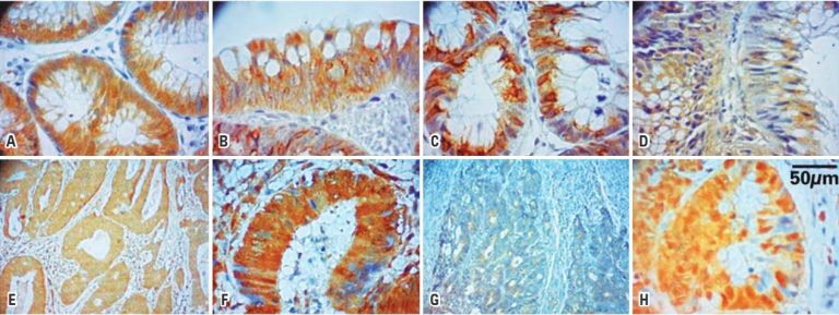
ABSTRACT Objective To evaluate the destruction complex of beta-catenin by the expression of the proteins beta-catetenin, adenomatous polyposis coli, GSK3β, axin and ubiquitin in colorectal carcinoma and colonic adenoma. Methods Tissue samples from 64 patients with colorectal carcinoma and 53 patients with colonic adenoma were analyzed. Tissue microarray blocks and slides were prepared and subjected to immunohistochemistry with polyclonal antibodies in carcinoma, adjacent non-neoplastic mucosa, and adenoma tissues. The immunoreactivity was evaluated by the percentage of positive stained cells and […]
Keywords: Adenoma; Axin protein; beta Catenin; Colorectal neoplasms; Genes, APC; Glycogen synthase; Immunohistochemistry; Ubiquitin; Wnt signaling pathway
31/Jan/2014
31/Jan/2014
DOI: 10.1590/S1679-45082013000400009
OBJECTIVE: To analyze the immunoexpression of the COX-2, p53, and caspase-3 proteins in colorectal adenomas and non-neoplastic mucosa. METHODS: 72 individuals were subjected to colonoscopy, which provided 50 samples of adenomas and 45 samples of non-neoplastic colorectal mucosa. The tissue samples were obtained via the tissue microarray technique and subjected to immunohistochemical analysis using primary anti-p53, anti-COX-2, and anti-caspase-3 antibodies. The positivity and intensity of the immunoreaction were classified. The analyzed variables were as follows: site of the adenomas in […]
Keywords: Adenoma; Caspase 3; Cyclooxygenase 2; Gene, p53; Immunohistochemistry; Intestine, large