einstein (São Paulo). 02/Feb/2026;24:eAI0595.
The Double ‘V’ sign of endomyocardial fibrosis
DOI: 10.31744/einstein_journal/2026AI0595
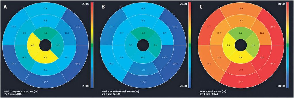
A 65-year-old woman presented to the cardiology department with exertional dyspnea and palpitations. Physical examination revealed bilateral jugular vein distension and mild lower-extremity edema. Blood tests indicated high B-type natriuretic peptide levels (777ng/mL). Echocardiography showed myocardial hypertrophy of the apical segments of both ventricles with cavity obliteration and left ventricular dysfunction with diffuse hypokinesia. Cardiovascular magnetic resonance (CMR) was performed to confirm the diagnosis. Cine images revealed biatrial enlargement, biventricular systolic dysfunction, and obliteration of both ventricular apical regions (, Video 1: ). Myocardial tissue characterization with late gadolinium enhancement (LGE) showed subendocardial enhancement of both ventricular apices (arrows in ) and overlying thrombus (symbol (*) in ) of the left ventricle (double “V” sign). Myocardial strain evaluated using the feature-tracking technique revealed an apicobasal gradient with reduced mobility in the apical segments. Although some basal segments did not show completely normal strain compared to the reference values, myocardial strain was significantly more impaired in the apical region. This highlighted the described gradient with greater impairment in the apical region.() (, Video 2: ). Cardiovascular magnetic resonance findings were consistent with those of endomyocardial fibrosis (EMF).
Endomyocardial fibrosis is a major cause of restrictive cardiomyopathy in tropical regions, particularly in underdeveloped countries.() Although the exact etiology is not well known, the disease is characterized by fibrotic tissue deposition in the endocardium of one or both ventricular apices.()
[…]
46