The journal einstein (São Paulo) – e-ISSN 2317-6385 is dedicated to dissemination of high-quality scientific content that advances our understanding of human disease with the goal of improving prevention, care, diagnostics and treatment of patients globally.
Technical advances in magnetic resonance imaging have allowed to accurately detect and grade endolymphatic space distension in Ménière disease; this was only possible in post-mortem histological studies until a few years ago. Magnetic resonance imaging rules out other causes of vertigo and hearing loss, and is able to evaluate the cochlear and vestibular compartments of the endolymphatic space using a dedicated protocol.
The role of magnetic resonance imaging in Ménière disease: the current state of endolymphatic hydrops evaluation
LoureiroRM, SumiDV, LemosMD, TamesHLVC, GomesRLE, DanielMM, SoaresCR, et al. The role of magnetic resonance imaging in Ménière disease: the current state of endolymphatic hydrops evaluation. Einstein (São Paulo) 2019;17(1):eMD4743. https://doi.org/10.31744/einstein_journal/2019MD4743
Loureiro,Rafael Maffei; Sumi,Daniel Vaccaro; Lemos,Marcelo Delboni; Tames,Hugo Luis de Vasconcelos Chambi; Gomes,Regina Lucia Elia; Daniel,Mauro Miguel; Soares,Carolina Ribeiro; Murakoshi,Rodrigo Watanabe; Funari,Marcelo Buarque de Gusmão. The role of magnetic resonance imaging in Ménière disease: the current state of endolymphatic hydrops evaluation. Einstein (São Paulo), v. 17, n. 1, eMD4743, Feb. 2019. https://doi.org/10.31744/einstein_journal/2019MD4743
Loureiro,R.M., Sumi,D.V., Lemos,M.D., Tames,H.L.V.C., Gomes,R.L.E., Daniel,M.M., Soares,C.R., Murakoshi,R.W., & Funari,M.B.G. (2019). The role of magnetic resonance imaging in Ménière disease: the current state of endolymphatic hydrops evaluation. Einstein (São Paulo),17(1), eMD4743. https://doi.org/10.31744/einstein_journal/2019MD4743
Loureiro,Rafael Maffei and Sumi,Daniel Vaccaro and Lemos,Marcelo Delboni and Tames,Hugo Luis de Vasconcelos Chambi and Gomes,Regina Lucia Elia and Daniel,Mauro Miguel and Soares,Carolina Ribeiro and Murakoshi,Rodrigo Watanabe and Funari,Marcelo Buarque de Gusmão. The role of magnetic resonance imaging in Ménière disease: the current state of endolymphatic hydrops evaluation. Einstein (São Paulo) [online]. 2019, vol. 17, n. 1, [cited 2026-06-13], eMD4743. Available from: <https://journal.einstein.br/article/the-role-of-magnetic-resonance-imaging-in-meniere-disease-the-current-state-of-endolymphatic-hydrops-evaluation/>. ISSN 1679-4508. https://doi.org/10.31744/einstein_journal/2019MD4743
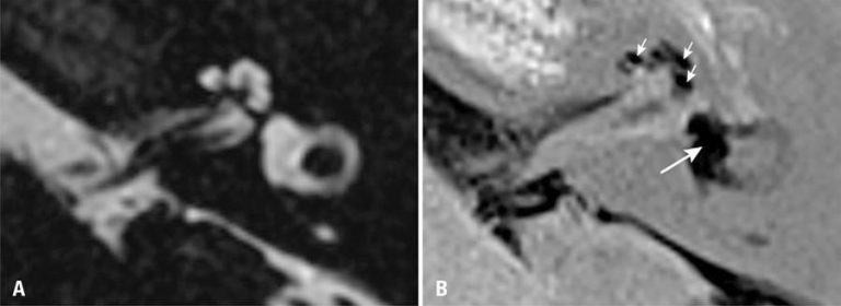
Figure 1
Axial magnetic resonance imaging of the left ear of a patient diagnosed with Ménière disease. (A) Highly T2-weighted sequence shows the labyrinthine fluid space (sum of endolymphatic and perilymphatic spaces). (B) Inversion recovery turbo spin echo with real reconstruction (3D real-IR) sequence 4 hours after intravenous gadolinium administration shows distension of saccule and utricle, which occupy most of the vestibular area (long arrow), and significant distension of cochlear duct (short arrows)
Axial magnetic resonance imaging of the left ear of a patient diagnosed with Ménière disease. (A) Highly T2-weighted sequence shows the labyrinthine fluid space (sum of endolymphatic and perilymphatic spaces). (B) Inversion recovery turbo spin echo with real reconstruction (3D real-IR) sequence 4 hours after intravenous gadolinium administration shows distension of saccule and utricle, which occupy most of the vestibular area (long arrow), and significant distension of cochlear duct (short arrows)