26/nov/2024
APENDIC-RADS: an ultrasound reporting system for the diagnosis of acute appendicitis
DOI: 10.31744/einstein_journal/2024AO1164
Highlights ■ Imaging plays a fundamental role in the diagnosis of acute appendicitis. ■ The study highlights ultrasound’s effectiveness in diagnosing appendicitis. ■ APPENDIC-RADS efficiently standardizes the ultrasound evaluation of appendicitis. ■ Using APPENDIC-RADS may enhance communication and optimize medical practices. ABSTRACT Objective: Abdominal ultrasonography is widely used to evaluate suspected cases of appendicitis. Objective descriptions of the direct and indirect signs of appendicitis result in varied assessments of its likelihood. This study introduces the Appendix Imaging Reporting and Data […]
Palavras-chave: Apendicite; Medicina de emergência; Comunicação interdisciplinar; Ultrassonografia
21/nov/2024
Point-of-care ultrasonography for diagnosis of purulent pericarditis postappendectomy: a case report
DOI: 10.31744/einstein_journal/2024RC0962
ABSTRACT Purulent pericarditis is rare condition in the modern era of antibiotics. However, it is a serious condition as it has an accelerated progression and is difficult to diagnose due to its nonspecific clinical presentation, resulting in high mortality. Herein, we present a case in which a 36-year-old male patient with otherwise unremarkable medical history developed abdominal sepsis complicated by purulent pericarditis post-appendectomy. While the initial clinical presentation was not compatible with the classic signs of purulent pericarditis, the diagnosis […]
Palavras-chave: Apendicectomia; Cuidados críticos; Pericardial effusion; Pericardite; Suppuration; Ultrassonografia
20/maio/2024
Lessons from the pandemic and the value of a structured system of ultrasonographic findings in the diagnosis of COVID-19 pulmonary manifestations
einstein (São Paulo). 20/maio/2024;22:eAE0780.
Ver Artigo20/maio/2024
Lessons from the pandemic and the value of a structured system of ultrasonographic findings in the diagnosis of COVID-19 pulmonary manifestations
DOI: 10.31744/einstein_journal/2024AE0780
Highlights Lung ultrasound enables precise diagnosis and ongoing monitoring of the disease. Ultrasound is an effective tool for assessing pulmonary findings in COVID-19. Structured reports enhance communication and are easily reproducible. ABSTRACT Objective: We aimed to share our experience in implementing a structured system for COVID-19 lung findings, elucidating key aspects of the lung ultrasound score to facilitate its standardized clinical use beyond the pandemic scenario. Methods: Using a scoring system to classify the extent of lung involvement, we retrospectively […]
Palavras-chave: Tomada de decisão clínica; Infecções por coronavírus; COVID-19; Hemossiderose; Organ dysfunction; SARS-CoV-2; Ultrassonografia
09/nov/2022
Sonographic signs of deep infiltrative endometriosis among women submitted to routine transvaginal sonography: clinical and imaging aspects
DOI: 10.31744/einstein_journal/2022AO0086
ABSTRACT Objective To evaluate the prevalence of sonographic signs suggestive of deep infiltrative endometriosis and endometriomas in patients referred for transvaginal sonography as part of a routine annual gynecological evaluation. We also describe the clinical and imaging aspects associated with the incidental findings of endometriosis. Methods This was a retrospective observational study including women (n=339; aged 18-56 years) referred for transvaginal sonography as part of a routine gynecological evaluation (without clinical suspicion of endometriosis). Patients were asked about their symptoms. […]
Palavras-chave: Neoplasias do endométrio; Endometriose; Prevalência; Ultrassonografia
28/jun/2022
Ultrasonographic evaluation of cecal appendix diameter in pediatric population
DOI: 10.31744/einstein_journal/2022AO6935
ABSTRACT Objective To stratify ultrasound samples in a pediatric population undergoing evaluation for acute appendicitis to examine the variability in cecal appendix diameter, in different age groups, and to determine whether there is a prevalent value for each age group. Methods A retrospective cross-sectional study with 196 children aged 0 to 15 years. Data were extracted from reports of ultrasound examinations carried out between 2008 and 2015. Children with sonographic diagnosis of appendicitis or other signs of periappendiceal inflammation were […]
Palavras-chave: Appendicitis/diagnostic imaging; Apêndice; Child; Ultrassonografia
06/maio/2022
Ultrasound classification of thyroid nodules: does size matter?
DOI: 10.31744/einstein_journal/2022AO6747
ABSTRACT Objective To determine whether the size of thyroid nodules in ACR-TIRADS ultrasound categories 3 and 4 is correlated with the Bethesda cytopathology classification. Methods Thyroid nodules (566) subclassified as ACR-TIRADS 3 or 4 were divided into three size categories according to American Thyroid Association guidelines. The frequency of different Bethesda categories in each size range within ACR-TIRADS 3 and 4 classifications was analyzed. Results Most nodules in both ACR-TIRADS classifications fell in the Bethesda 2 category, regardless of size […]
Palavras-chave: ACR-TIRADS; Bethesda; Malignance; Thyroid gland nodule; Thyroid gland/pathology; Ultrassonografia
07/abr/2022
Testicular volume and Tanner stage: determinant factors for testicular torsion
DOI: 10.31744/einstein_journal/2022AO6605
ABSTRACT Objective To assess testicular volumes and sexual maturation in patients with testicular torsion. Methods A retrospective analysis of consecutively treated patients with testicular torsion between 2016 and 2018. Age, pubic hair staging (Tanner), and by ultrasonography, volume of the unaffected testis (in cubic centimeters) were evaluated either immediately before surgery or at the first postoperative visit. Patients with previous testicular disease, such as cryptorchidism, or with no records of testicular volume were excluded. The analysis included descriptive statistics and […]
Palavras-chave: Isquemia; Sexual maturation; Torção do cordão espermático; Testis/diagnostic imaging; Testis/growth & development; Ultrassonografia
26/nov/2020
Lipomatose do apêndice cecal em paciente grávida mimetizando apendicite aguda
DOI: 10.31744/einstein_journal/2020RC5415
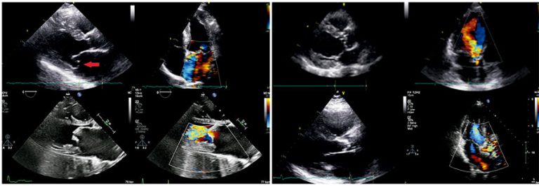
RESUMO Paciente de 34 anos, do sexo feminino, grávida, chega ao pronto-socorro com queixa de dor na fossa ilíaca direita piorando nos últimos 2 dias com suspeita de apendicite aguda. Foram solicitados exames laboratoriais, que estavam dentro dos limites de normalidade para aspectos infecciosos e inflamatórios. Exame de imagem também foi solicitado, sendo a ultrassonografia o método de escolha, que revelou gravidez em curso sem alterações e espessura da parede do apêndice sem sinais inflamatórios. Ainda com suspeita de apendicite […]
Palavras-chave: Apendicite; Diagnóstico diferencial; Gestantes; Imagem por ressonância magnética; Lipomatose; Ultrassonografia
06/nov/2020
Estudo comparativo de ultrassonografia e métodos seccionais para detecção de pequenas lesões renais: fatores anatômicos e experiência do radiologista
einstein (São Paulo). 06/nov/2020;18:eAO5576.
Ver Artigo06/nov/2020
Estudo comparativo de ultrassonografia e métodos seccionais para detecção de pequenas lesões renais: fatores anatômicos e experiência do radiologista
DOI: 10.31744/einstein_journal/2020AO5576
RESUMO Objetivo: Avaliar os fatores anatômicos e a experiência do radiologista na detecção de massas renais sólidas na ultrassonografia. Métodos: Buscamos massas renais sólidas diagnosticadas em imagens seccionais, de 2007 a 2017, que também tivessem ultrassonografia prévia nos últimos 6 meses. As seguintes características foram avaliadas: tamanho do nódulo, lateralidade, localização e padrão de crescimento, índice de massa corporal do paciente e experiência do radiologista em ultrassonografia. Nos casos com ressecção cirúrgica, os laudos de patologia foram analisados. O teste […]
Palavras-chave: Diagnóstico por imagem; Neoplasias renais/diagnóstico por imagem; Tomografia computadorizada multidetectores; Ultrassonografia
28/jan/2020
Avaliação do volume prostático obtido por ressonância magnética multiparamétrica da próstata e ultrassonografia transretal comparado ao peso da peça cirúrgica
DOI: 10.31744/einstein_journal/2020AO4662
RESUMO Objetivo Avaliar a acurácia da ressonância magnética multiparamétrica da próstata para estimativa do volume da glândula, comparando seus resultados com a ultrassonografia transretal e correlacionando com o volume obtido da peça cirúrgica. Métodos Estudo retrospectivo incluindo 85 pacientes submetidos à ressonância magnética e, posteriormente, à ultrassonografia transretal (para orientação de biópsia com fusão de imagens) e, a seguir, à prostatectomia radical. As dimensões prostáticas foram obtidas na ressonância a partir das imagens nos planos axial e sagital em sequências […]
Palavras-chave: Doenças prostáticas; Espectroscopia de ressonância magnética; Imagem por ressonância magnética; Prostatectomia; Ultrassonografia