einstein (São Paulo). 04/Jul/2016;14(3):435-6.
Spontaneous coronary artery dissection and healing documented by optical coherence tomography
DOI: 10.1590/S1679-45082016AI3551
An otherwise healthy 57-year-old female patient with no risk factors for coronary artery disease presented to the emergency room with acute chest pain. The patient had been taking appetite suppressant (dimethylamylamine, Oxyelite Pro, (USP Labs) for the last 7 days.,
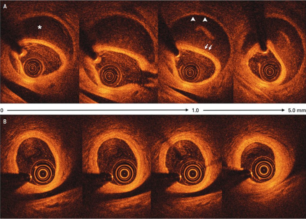
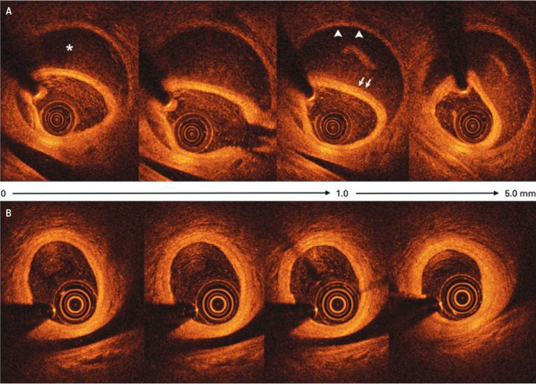
Electrocardiography showed no abnormalities although both serum creatinine kinase-MB (3.59ng/mL) and troponin I (8,310pg/mL) were elevated. Coronary angiography revealed extensive and abrupt lumen narrowing in the obtuse marginal with a subtle intraluminal defect within the distal part of the vessel (). Optical coherence tomography (OCT), which is a novel near-infrared light based intravascular imaging modality with high-resolution images (10 to 20μm), showed an intramural hematoma that was 20mm long, with a 10mm length of near-circumferential dissection (double lumen) () with no evidence of atherosclerosis. The patient was discharged after medical management with aspirin, clopidogrel, low-molecular weight heparin. Six months later a new coronary angiography and OCT were performed revealing a complete spontaneous resolution of the dissection ( and ).
[…]
253