The journal einstein (São Paulo) – e-ISSN 2317-6385 is dedicated to dissemination of high-quality scientific content that advances our understanding of human disease with the goal of improving prevention, care, diagnostics and treatment of patients globally.
A 45-year-old male patient, hospitalized and under investigation of mediastinal mass, presenting with sudoresis, malaise, and dysphagia. Computed tomography showed eccentric parietal thickening of the middle third of esophagus, with mass effect and upstream ectasia, with no expressive increase of metabolic activity. Additionally, there are cervical, axillary, pulmonary hilar, portocaval space, and unspecific iliac lymph nodes ( and ).
ReimãoSM, ColaiacovoR, CamunhaMAR, AmancioTT, SegatelliV, PauloGA. Mediastinal tumor: not always a lymphoma. Einstein (São Paulo) 2017;15(3):1-2. https://doi.org/10.1590/S1679-45082017AI3981
Reimão,Silvia Mansur; Colaiacovo,Rogério; Camunha,Marco Antonio Ribeiro; Amancio,Thiago Trolez; Segatelli,Vanderlei; Paulo,Gustavo Andrade de. Mediastinal tumor: not always a lymphoma. Einstein (São Paulo), v. 15, n. 3, p. 1-2, Jul. 2017. https://doi.org/10.1590/S1679-45082017AI3981
Reimão,S.M., Colaiacovo,R., Camunha,M.A.R., Amancio,T.T., Segatelli,V., & Paulo,G.A. (2017). Mediastinal tumor: not always a lymphoma. Einstein (São Paulo),15(3), 1-2. https://doi.org/10.1590/S1679-45082017AI3981
Reimão,Silvia Mansur and Colaiacovo,Rogério and Camunha,Marco Antonio Ribeiro and Amancio,Thiago Trolez and Segatelli,Vanderlei and Paulo,Gustavo Andrade de. Mediastinal tumor: not always a lymphoma. Einstein (São Paulo) [online]. 2017, vol. 15, n. 3, [cited 2026-02-19], pp.1-2. Available from: <https://journal.einstein.br/article/mediastinal-tumor-not-always-a-lymphoma/>. ISSN 1679-4508. https://doi.org/10.1590/S1679-45082017AI3981
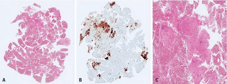
Figure 4
(A) Slide of the ultrasound-guided biopsy specimen (4x, hematoxylin and eosin). (B) Cells showing positive immunoexpression for desmin. (C) Magnified area showing bundles of smooth muscle cells (20x, hematoxylin and eosin)
(A) Slide of the ultrasound-guided biopsy specimen (4x, hematoxylin and eosin). (B) Cells showing positive immunoexpression for desmin. (C) Magnified area showing bundles of smooth muscle cells (20x, hematoxylin and eosin)