einstein (São Paulo). 01/Jan/2016;14(1):106-7.
Giant ulcerative lesion on the upper back: using a differential diagnosis to formulate a clinical approach
DOI: 10.1590/S1679-45082016AI3405
A 57-year-old white man with no significant past medical history presented to the county hospital emergency room with complaints of increasing fatigue and lightheadedness over the past year. Upon further questioning, he revealed a large ulcerative lesion on his upper back that he reported started as a small ulcer and progressed to its current size over a 16-year period. The patient had not sought any medical attention throughout this time.
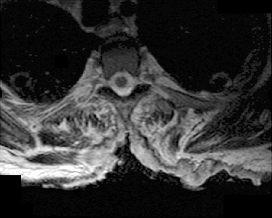
The patient had no known history of malignancy, immunosuppressive conditions, autoimmune disorders, exposure to communicable diseases, or travel outside of the United States. His vital signs were within normal limits. Physical examination revealed a 26cmx16cm ulcerative lesion spanning the T1 through T8 vertebral bodies with exposure of the spinous processes and paravertebral musculature, which was most prominent at the level of T5. The lesion contained punctate areas of bleeding, granulation tissue, and copious serous drainage. The boarders were clearly defined and without satellite lesions (). Other than pallor of the skin, the remainder of the physical examination, including a full neurological assessment, was unremarkable. In the emergency room, a computed tomography scan of the chest/abdomen/pelvis was performed and two individual punch biopsies of the ulcer bed were taken. The computed tomography scan showed erosion of the thoracic spinous processes but no evidence of metastatic disease. A complete blood count revealed a hemoglobin and white blood count of 4.6g/dL and 6.9 cells x 103/µL, respectively. On admission the patient was transfused for his symptomatic anemia and started on ferrous sulfate.
[…]
194