08/May/2019
Vertebral body erosion secondary to aortoiliac aneurysm
DOI: 10.31744/einstein_journal/2019AI4550
A 75-year old female patient presenting chronic arterial hypertension, was admitted at the emergency department with moderate unspecific abdominal pain. A computed tomography of total abdomen with no contrast enhancement was requested due to altered renal function and suspicion of acute obstructive abdomen. The computed tomography sections ( and ) showed a fusiform aneurysm in the aortoiliac bifurcation, extending up to the right iliac artery. This aneurysm was causing erosion of the anterior aspect of L4 vertebral body, with bone […]
02/May/2019
Pneumopericardium: a rare case of cardiorespiratory arrest
DOI: 10.31744/einstein_journal/2019AI4516
A 37-days-old baby boy, previously healthy and without intercurrences during perinatal period, who was admitted in pediatric intensive care unit with acute ventilator insufficiency because of viral bronchiolitis due to respiratory syncytial virus. The patient underwent a tracheal intubation by clinical deterioration, a rapid sequence intubation using fentanyl, ketamine and succinylcholine. During the procedure, he had cough, chest rigidity, reduction of respiratory sounds and poor tissue perfusion. His clinical picture worsened and resulted in cardiorespiratory arrest in asystole that was […]
28/Feb/2019
Multiple laryngeal neuromas: rare benign tumor
DOI: 10.31744/einstein_journal/2019AI4465
A 66-year old female patient complained of sensation of a foreign body in her pharynx and of dry cough for the past 6 months. She denied dysphonia, dysphagia or dyspnea. Endoscopy was performed for suspected gastroesophageal reflux, and it showed several roundish, yellowish and well-limited lesions, in the posterior region of the larynx, adjacent to the arytenoid cartilages ( and ). A biopsy of the lesions was performed ( ), and the pathological examination revealed neoplasm with clear limits, composed […]
25/Feb/2019
Congenital nasal pyriform aperture stenosis with single central megaincisor
DOI: 10.31744/einstein_journal/2019AI4525
A 15-day-old female newborn presenting nasal obstruction since birth underwent computed tomography of the paranasal sinuses in our department due to clinical suspicion of choanal atresia. The patient presented no other complaints. Computed tomography () allowed the diagnosis of pyriform aperture stenosis, which is a rare cause of nasal obstruction in neonates and was first described in 1989.() Pyriform aperture stenosis becomes particularly significant during the first two months of life when infants are mandatory nose breathers. […]
25/Feb/2019
Idiopathic masseter muscle hypertrophy: a case report
DOI: 10.31744/einstein_journal/2019AI4506
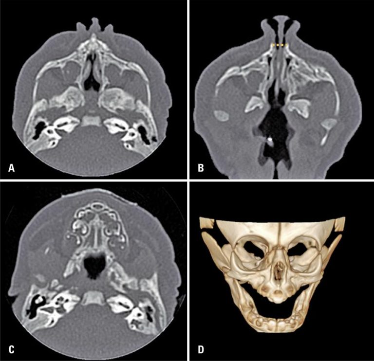
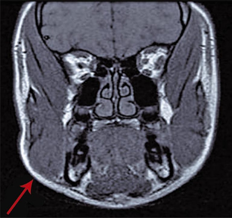
Idiopathic masseter muscle hypertrophy is a rare condition, characterized by unilateral or bilateral enlargement of this muscle, sometimes associated to mandibular angle exostosis.() Its etiology is unknown, and there is a possible relation with unilateral masticatory activity, dental malocclusion, temporomandibular joint dysfunction, bruxism, or emotional alterations.(,) The diagnosis is made primarily based on the clinical presentation, complemented with ultrasound and, if required, magnetic resonance imaging.() It is important to make differential diagnoses with tumors or inflammatory processes in muscles, bones […]
28/Jan/2019
Sellar and suprasellar arachnoid cyst
DOI: 10.31744/einstein_journal/2019AI4269
A 36-year-old woman admitted with a 2-week history of headaches and blurred vision. Her medical history was positive for irregular menses and hypothyroidism. Visual field tests revealed defects in the upper quadrants bilaterally and blood tests indicated slightly elevated prolactin levels (24.4; range 4.8 to 23.3), reduced morning cortisol (3.8; range 4 to 22), and reduced growth hormone levels (
12/Dec/2018
Challenge in the diagnosis of pulmonary strongyloidiasis
DOI: 10.31744/einstein_journal/2019AI4441
A 33-years-old man was admitted to the intensive care unit because of alcoholic cirrhosis, cocaine toxicity, and septic shock to be accurately assessed. Further tests were requested. His X-ray did not show compatible patterns with pulmonary strongyloidiasis, blood culture was positive for enterobacteria that produce Klebsiella pneumoniae carbapenemase (KPC), and discrete eosinophylin hemogram and tracheal secretion culture were observed. […]
30/Oct/2018
Isolated middle colic artery injury after blunt abdominal trauma
DOI: 10.31744/einstein_journal/2018AI4384
A 34-year old male patient, victim of motorcycle accident at about 56 miles/hour, was brought into emergency care with a cervical collar. He was hemodynamically stable, with no respiratory or neurological impairment, complaining of diffuse moderate intensity abdominal pain during the physical examination. During secondary evaluation, abrasions were identified on the abdominal wall in the right flank area. Due to high energy trauma, he underwent a whole body computed tomography (CT) that showed an hematoma in the mesenteric fat planes […]
10/Sep/2018
Marsupial vena cava mimicking lymph node enlargement on tomography
DOI: 10.1590/S1679-45082018AI4394
A 56-year-old male patient, hypertensive, diabetic and asymptomatic, underwent abdominal computed tomography (CT) with intravenous contrast for follow-up of hepatic steatosis. Computed tomography demonstrated a solid hypervascular nodule in the pancreatic head (characteristics of neuroendocrine lesion – ) and oval formation anteriorly to the aortoiliac bifurcation (). In this situation, axial CT can mimick lymph node enlargement, especially in clinical oncology context. However, evaluation of the different phases of the exam and the reformatted coronal and sagittal images help making […]
05/Jun/2018
Atypical hepatic hemangioma: imaging features of hyalinized hemangioma
DOI: 10.1590/S1679-45082018AI4256
A 46 years old man, complaining about abdominal pain, was submmited to a magnetic resonance having a giant liver hemangioma as an incidental finding (). In the follow-up, after 6 years, we observed in a T2-weighted sequence volumetric reduction of lesions’ signal in T2-weighted image () was observed. Temporal evolution, associated with imaging features, indicated sclerosed/hyalinized hemangioma (). […]
