3 results
23/Oct/2025
23/Oct/2025
DOI: 10.31744/einstein_journal/2025AO1592
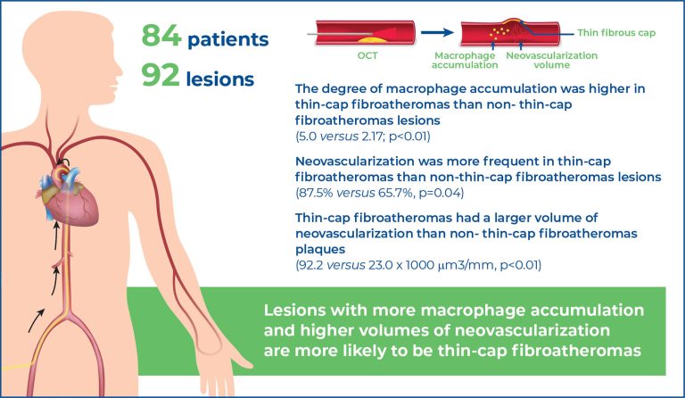
Highlights ■ Optical coherence tomography enables in vivo characterization of inflammatory activity within coronary plaques. ■ Thin-cap fibroatheromas demonstrate greater macrophage accumulation than non-thin-cap fibroatheromas. ■ Neovascularization is more frequent and quantitatively greater in thin-cap fibroatheromas. ■ The combined burden of macrophages and neovessels provides predictive value for plaque vulnerability. ABSTRACT Objective: The aim of the present study is to assess whether the intensity of local inflammation relates to the presence of thin-cap fibroatheromas. Methods: Retrospective, single-center study of patients […]
Keywords: Atherosclerosis; Coronary artery disease; Inflammation; Microphages; Plaque, atherosclerotic; Tomography, optical coherence
19/Feb/2021
DOI: 10.31744/einstein_journal/2021RC5521
ABSTRACT Sophisticated imaging systems have helped to redefine the clinical presentation of acute macular neuroretinopathy and have markedly enhanced diagnostic sensitivity. The proposed mechanism of paracentral acute middle maculopathy is related to ischemia at the level of the superficial and deep retinal capillary plexi. This is a case report of a patient who developed an acute macular neuroretinopathy after an uneventful angioplasty with stents in the coronary artery.
Keywords: Angioplasty; Constriction, pathologic; Macula lutea; Macular degeneration; Retinal vessels; Stents; Tomography, optical coherence
01/Jan/2014
DOI: 10.1590/S1679-45082014RC2708
We report the case of a patient with congenital toxoplasmosis and submacular hemorrhage caused by a neovascular membrane who underwent an intravitreal injection of C3F8 and bevacizumab, and had a good visual recovery.
Keywords: Angiogenesis inhibitors; Case reports; Intravitreal injections/adverse effects; Retinal hemorrhage/etiology; Retinal neovascularization/complications; Tomography, optical coherence; Toxoplasmosis, congenital/etiology