The journal einstein (São Paulo) – e-ISSN 2317-6385 is dedicated to dissemination of high-quality scientific content that advances our understanding of human disease with the goal of improving prevention, care, diagnostics and treatment of patients globally.
Appendiceal diverticulitis is an uncommon condition, mimicking appendicitis, but with greater risk of perforation and complications. Preoperative diagnosis is rare, but can be achieved by ultrasonography as identification of the diverticulum and classical signs of appendicitis. We report a case of ultrasonographic diagnosis of a perforated appendiceal diverticulitis in an adult male and discuss this condition.
Perforated diverticulitis of the appendix: ultrasonographic diagnosis
LourençoRB, PinhoMC, SchraibmanV, MacedoALV, Francisco NetoMJ, FunariMBG. Perforated diverticulitis of the appendix: ultrasonographic diagnosis. Einstein (São Paulo) 2011;9(1 Pt 1):75-7. https://doi.org/10.1590/S1679-45082011RC978
Lourenço,Rafael Burgomeister; Pinho,Marco da Cunha; Schraibman,Vladimir; Macedo,Antônio Luiz de Vasconcellos; Francisco Neto,Miguel José; Funari,Marcelo Buarque de Gusmão. Perforated diverticulitis of the appendix: ultrasonographic diagnosis. Einstein (São Paulo), v. 9, n. 1 Pt 1, p. 75-77, Jan. 2011. https://doi.org/10.1590/S1679-45082011RC978
Lourenço,R.B., Pinho,M.C., Schraibman,V., Macedo,A.L.V., Francisco Neto,M.J., & Funari,M.B.G. (2011). Perforated diverticulitis of the appendix: ultrasonographic diagnosis. Einstein (São Paulo),9(1 Pt 1), 75-77. https://doi.org/10.1590/S1679-45082011RC978
Lourenço,Rafael Burgomeister and Pinho,Marco da Cunha and Schraibman,Vladimir and Macedo,Antônio Luiz de Vasconcellos and Francisco Neto,Miguel José and Funari,Marcelo Buarque de Gusmão. Perforated diverticulitis of the appendix: ultrasonographic diagnosis. Einstein (São Paulo) [online]. 2011, vol. 9, n. 1 Pt 1, [cited 2026-02-23], pp.75-77. Available from: <https://journal.einstein.br/article/perforated-diverticulitis-of-the-appendix-ultrasonographic-diagnosis/>. ISSN 1679-4508. https://doi.org/10.1590/S1679-45082011RC978
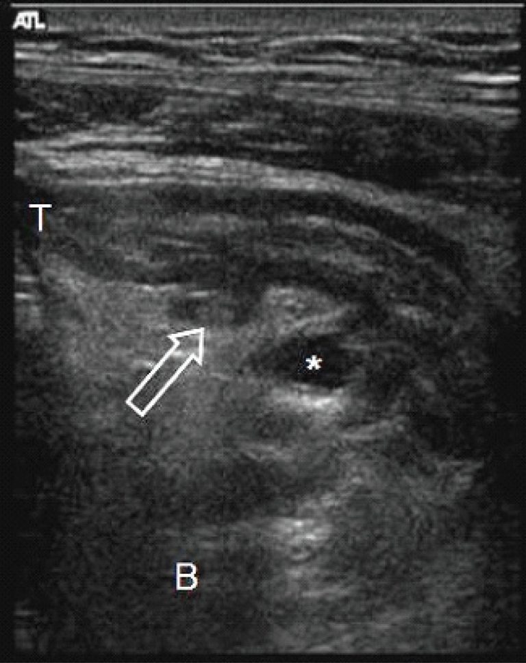
Figure 2
Localized parasagittal image. Small fluid collection (*) adjacent the perforated diverticulum. Another non-perforated diverticulum (open arrow) was identified near the tip (T). B: base.
Localized parasagittal image. Small fluid collection (*) adjacent the perforated diverticulum. Another non-perforated diverticulum (open arrow) was identified near the tip (T). B: base.