86 results
07/Nov/2023
07/Nov/2023
DOI: 10.31744/einstein_journal/2023AI0386
A 49-year-old woman with a history of pulmonary thromboembolism and right ventricular dysfunction sought consultation with a pulmonologist at our hospital, reporting exertion dyspnea and four episodes of syncope with prodromal palpitations. Physical examination results were unremarkable, and the patient had no history of heart disease. Cardiac magnetic resonance imaging was performed for proper evaluation of the right ventricle. Cardiac magnetic resonance showed normal biventricular function; however, tricuspid valve prolapse and tricuspid annulus disjunction were observed (maximum separation between the […]
30/Oct/2023
30/Oct/2023
DOI: 10.31744/einstein_journal/2023AI0538
The use of herbal medicines is growing exponentially despite the lack of scientific evidence regarding their effectivity and toxicity. ( , ) Moreover, the use of herbal medicines have been reported to be common in patients with chronic liver disease (30–62%); these medicines can lead to hepatotoxicity and serious hepatic side effects. ( ) Hepatic encephalopathy is a complication of acute liver failure and chronic liver disease that causes cognitive dysfunction, motor deficits, and seizures. ( , ) Gastrointestinal hemorrhage, […]
12/Aug/2022
DOI: 10.31744/einstein_journal/2022AI0008
Umbilical cord haematoma (UCH) is a spontaneous bleeding in the umbilical cord caused by the rupture of umbilical vessels with blood extravasation in the Warton’s jelly. This condition is a rare complication of pregnancy (1:5000-11000 deliveries). UCH may cause fetal distress and it can be associated with stillbirth (50% of cases). For these reasons, close surveillance is required when diagnosed by antenatal ultrasound.( – ) Various risk factors have been suggested such as cord anomalies, infection, coagulation disorder, post maturity, […]
10/Aug/2022
DOI: 10.31744/einstein_journal/2022AI6853
Left ventricular noncompaction (LVNC) is a relatively new and heterogeneous cardiomyopathy, first reported by Chin et al. in 1990.() It is characterized by prominent ventricular trabeculations, intratrabecular recesses and a bi-layered myocardium, composed of compacted and noncompacted layers.(,) Left ventricular noncompaction may be an isolated finding or associated to other cardiomyopathies, metabolic disorders and congenital heart diseases.() It is believed to result from failure of ventricular compaction in the embryogenic phase, with increasing evidence of a genetic component.() Since its […]
13/Apr/2022
DOI: 10.31744/einstein_journal/2022AI6672
An 18-year-old female patient with hypertrophic cardiomyopathy previously asymptomatic, diagnosed in family screening, who presented progressive dyspnea even upon mild exertion two months before. She was submitted to cardiac magnetic resonance ( and ), which showed asymmetrical myocardial hypertrophy with mid-ventricular septal predominance. The most common form of obstruction of the left ventricle outflow tract in hypertrophic cardiomyopathy is subaortic obstruction,() which generally results from left ventricle outflow tract narrowing by septal hypertrophy and systolic anterior motion of the mitral […]
15/Dec/2021
DOI: 10.31744/einstein_journal/2021AI6683
A 34-year-old patient with a history of asthma, taking inhaled corticosteroids and bronchodilators, presented with worsening dyspnea (functional class III) and reports of syncope. On physical examination, pulmonary auscultation was normal with pulse oximetry of 96% in room air. On cardiac auscultation, the rhythm was regular with fixed splitting S2 and systolic murmur 2+/6+ on left sternal border. On abdominal examination, the liver was palpable 2cm below the right costal border. There were no other signs of heart failure. Chest […]
15/Dec/2021
DOI: 10.31744/einstein_journal/2021AI6347
A 15-year old women presented with a 6-month history of progressive right-hand tremor with functional impairment, aggravated by a 1-month history of episodic confusion. The patient underwent a computed tomography scan, which revealed a posterior fossa expansive lesion with cerebrospinal fluid-like density and a midline bone discontinuity ( ; soft tissue window not shown). A magnetic resonance imaging (MRI) scan ( ) was performed after hospital admission, revealing a posterior fossa expansile lesion with predominant T2 hyperintensity, T1 hypointensity, linear […]
03/Dec/2021
DOI: 10.31744/einstein_journal/2021AI6564
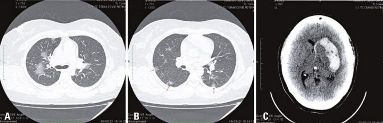
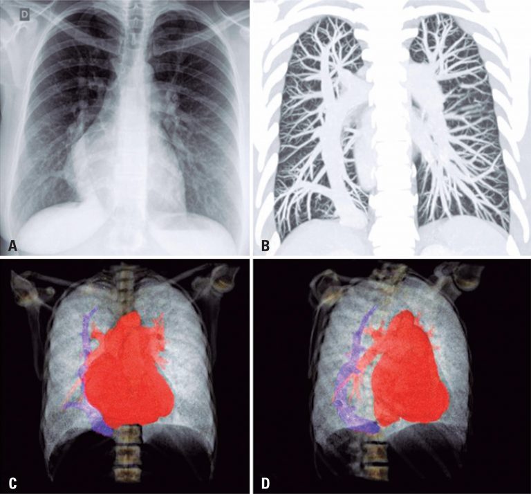
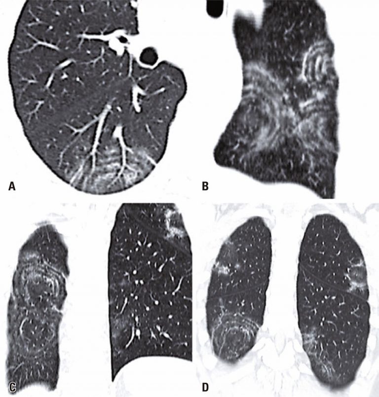
A 49-year-old man came to our emergency department with a 2-day history of fever, cough, anosmia, ageusia and odynophagia. His past medical history included hypertension. At the time of this presentation, chest computed tomography revealed peripheral and bilateral ground-glass opacities, with some visible intralobular lines – typical findings of pneumonia caused by the severe acute respiratory syndrome coronavirus 2 (SARS-CoV-2). In addition, some findings revealed the target sign (). The patient’s supportive treatment was continued, and reverse-transcriptase polymerase chain reaction […]
01/Apr/2021
DOI: 10.31744/einstein_journal/2021AI5707
Carotid artery atherosclerosis is one of the main causes of stroke.() In dental panoramic radiography (DPR), radiopaque images adjacent to cervical spine can be an indicative of calcification in bifurcation of carotid artery. Such calcifications can represent calcified atheroma plaques,() which diagnosis is confirmed by the Doppler ultrasonography.(,) Dentists can identify these areas through DPR and differentiate them from radiopacities that can be observed in the carotid region. The identification of these area and referral of these patients to specialized […]
27/Jan/2021
DOI: 10.31744/einstein_journal/2021AI5829
A 55-year-old man admitted to our emergency department complaining of right flank acute pain. He also referred epistaxis, gingival bleeding, and hematuria for the past 3 days. His physical exam demonstrated abdominal subcutaneous hematomas and he referred abdominal tenderness on palpation. Seven days before these symptoms, his daily warfarin dose has been increased. His blood tests showed that the International Normalized Ratio was markedly elevated (3.6; his previous exam from before dosage increase was 2.2). An abdominal computed tomography was […]