einstein (São Paulo). 06/maio/2024;22:eAO0764.
Assessing the toxicity of one-step-synthesized PEG-coated gold nanoparticles: in vitro and in vivo studies
DOI: 10.31744/einstein_journal/2024AO0764
Highlights
One-step synthesis provides affordable, stable, and biocompatible nanoparticles.
PEG-coated gold nanoparticles exhibited very low cytotoxicity effects.
This in vivo study did not reveal hematopoietic, renal, or hepatic alterations.
The histopathological analysis did not present any tissue or cellular damage.
ABSTRACT
Objective:
To evaluate the in vitro and in vivo toxicities of polyethylene glycol-coated gold nanoparticles synthesized using a one-step process.
Methods:
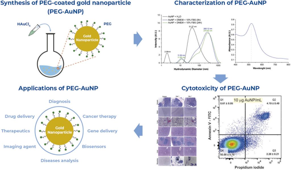
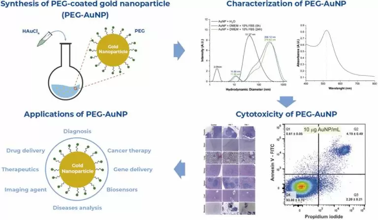
Gold nanoparticles were prepared via a co-precipitation method using polyethylene glycol, and the synthesis product was characterized. For the in vitro evaluation, a flow cytometry analysis with Annexin V and iodide propidium staining was used to assess cytotoxicity in MG-63 cells labeled with 10, 50, and 100μg/mL of nanoparticle concentration. For the in vivo evaluation, nanoparticles were administered intraperitoneally at a dose of 10mg/kg dose in 10-week-old mice. Toxicity was assessed 24 hours and 7 days after administration via histopathological analysis of various tissues, as well as through renal, hepatic, and hematopoietic evaluations.
Results:
Synthesized nanoparticles exhibited different hydrodynamic sizes depending on the medium: 51.27±1.62nm in water and 268.12±28.45nm (0 hour) in culture medium. They demonstrated a maximum absorbance at 520nm and a zeta potential of -8.419mV. Cellular viability exceeded 90%, with less than 3% early apoptosis, 6% late apoptosis, and 1% necrosis across all labeling conditions, indicating minimal cytotoxicity differences. Histopathological analysis highlighted the accumulation of nanoparticles in the mesentery; however, no lesions or visible agglomeration was observed in the remaining tissues. Renal, hepatic, and hematopoietic analyses showed no significant differences at any time point.
Conclusion:
Polyethylene glycol-coated gold nanoparticles exhibit extremely low toxicity and high biocompatibility, showing promise for future studies.
[…]
Palavras-chave: Nanomedicine; Flow cytometry; In vitro techniques; Nanoparticles; polyethylene glycols; Toxicity
630