einstein (São Paulo). 23/out/2025;23:eAO1592.
Thin-cap fibroatheroma association with local inflammatory activity in coronary disease: an optical-coherence tomography study
DOI: 10.31744/einstein_journal/2025AO1592
Highlights
■ Optical coherence tomography enables in vivo characterization of inflammatory activity within coronary plaques.
■ Thin-cap fibroatheromas demonstrate greater macrophage accumulation than non-thin-cap fibroatheromas.
■ Neovascularization is more frequent and quantitatively greater in thin-cap fibroatheromas.
■ The combined burden of macrophages and neovessels provides predictive value for plaque vulnerability.
ABSTRACT
Objective:
The aim of the present study is to assess whether the intensity of local inflammation relates to the presence of thin-cap fibroatheromas.
Methods:
Retrospective, single-center study of patients that underwent optical coherence tomography imaging and had either de novo or in-stent neoatherosclerosis. Intensity of macrophage accumulation and volume of neovascularization were measured for all lesions. Logistic binary regressions were used for uni- and multivariate analysis.
Results:
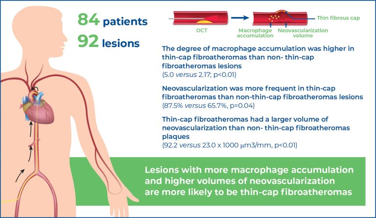
A total of the 92 lesions in 84 patients were selected. The degree of macrophage accumulation was higher in thin-cap fibroatheromas than non- thin-cap fibroatheromas lesions (5.0 versus 2.17; p<0.01). Neovascularization was more frequent in thin-cap fibroatheromas than non-thin-cap fibroatheromas lesions (87.5% versus 65.7%, p=0.04), and thin-cap fibroatheromas had a larger volume of neovascularization than non- thin-cap fibroatheromas plaques (92.2 versus 23.0 x 1000μm3/mm, p<0.01). At multivariate logistic analysis, neovascularization volume and degree of macrophage accumulation remained independently associated with thin-cap fibroatheromas. The dataset was divided according to the highest tercile of neovascularization volume (≥87.2 x 1000μm3/mm) and macrophage accumulation score (≥4.6). Plaques with low levels of neovascularization and macrophages were classified as thin-cap fibroatheromas in 14% of cases. Thin-cap fibroatheromas was present in 61.5% of plaques with high macrophagic and neovascularization content.
Conclusion:
Lesions with more macrophage accumulation and higher volumes of neovascularization are more likely to be thin-cap fibroatheromas.
[…]
67