einstein (São Paulo). 17/dez/2025;24:eRW0987.
Computed tomography imaging features of major pulmonary and extrapulmonary complications of fibrotic lung diseases
DOI: 10.31744/einstein_journal/2026RW0987
ABSTRACT
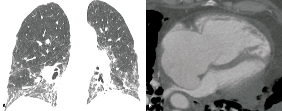
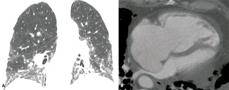
Patients diagnosed with fibrosing interstitial lung disease are at risk of several complications, some of which may be life-threatening. Computed tomography imaging plays an important role in diagnosing these overlapping conditions. This article summarizes the computed tomography imaging features of the most common conditions associated with fibrosing interstitial lung diseases, categorized by involvement of the lung parenchyma or extra-pulmonary compartments. Some steps may help to recognize such complications, such as having knowledge of the underlying fibrotic lung disease, being aware of potentially immunosuppressive treatments in use, noting new relevant symptoms, checking previous imaging examinations to detect subtle changes, and considering the exam technique, for example, to avoid false perception of ground-glass opacities in exams with insufficient inspiration. Unenhanced computed tomography may be enough to diagnose most situations, but in specific cases, for example, in the clinical suspicion of pulmonary embolism, dedicated computed tomography angiography may be warranted. Careful comparison with previous exams is advised, especially to detect subtle opacities suspicious for lung cancer, underscoring that its detection may be difficult owing to the baseline morphological lung changes. Radiologists must be aware of such possible complications to perform early diagnosis and ensure proper management.
63